
The Inner Workings of the Eye
Have you ever stopped to think about how amazing the human eye is?
These complicated organs take light waves bouncing off the things in front of us and convert those into a continuous stream moving images that are accurate and detailed. They arguably connect us to our surroundings more than any other sense and make so many activities we take for granted possible, from driving to reading to looking at our loved ones’ faces. But how do they do all that?
How the Parts of the Eye Help Us See
Let’s focus today on the portions of eye anatomy that are closely involved in creating the images that go to the brain. We’ll move from front to back.
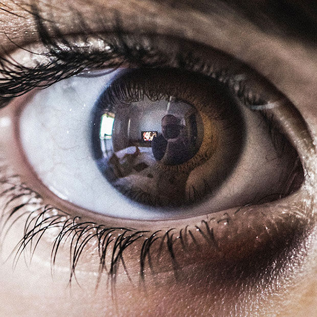
- Cornea: the clear layer at the very front of the eye that allows light inside.
- Iris: the colorful, circular muscle that contracts and expands to control the amount of light entering the eye.
- Lens: a clear disc that changes shape in order to focus on objects at different distances. (This is different from the lens in a camera, which is rigid and must physically move in order to change focus.)
- Retina: a thin layer of tissue at the back of the eye covered in light-sensitive cells called rods and cones.
- Optic nerve: the nerve that transmits visual information from the retina to the brain for processing at an estimated rate of around a million bits per second! That’s a lot!
Eyes Working As a Team: Binocular Vision
Have you ever noticed that you see a slightly different angle with your right eye than you do with your left? You can test it by covering one eye at a time. This is binocular vision. The differences in the two images creates a live 3D image. It’s the reason we have depth perception, or the ability to judge the relative distances of different objects from us.
The Visual Cortex of the Brain
The visual cortex is located in the occipital lobe in the back of the brain. About 20% of the brain is dedicated to visual processing, and another 40% is involved in smaller ways, like vision+touch, vision+motor, vision+attention, vision+meaning, etc. We’re absorbing and processing new visual data many times per second, which is how we can perceive motion, make sense of what we see, and react.
The Eyes’ Defense Team and Maintenance Crew
We’ve talked about the parts of the eye involved in turning light waves into images processed in the brain, but there are other parts whose job is to keep the whole system running smoothly. Our eyelids are there to protect our eyes. Eyebrows and eyelashes help with that. When we blink, it refreshes the tear film and sweeps away any debris and contaminants. The tear film itself is produced and maintained by a system of glands and ducts.
Help Your Eyes Stay in Good Shape
We will never stop being amazed by the way all the many parts of the eye work together to make vision possible. Being as complex as eyes are, though, there are quite a few ways things can go wrong if they aren’t getting the care they need. That’s where we come in! Make sure to let us know if you’ve noticed any recent changes in your vision or have been experiencing any eye-related symptoms.